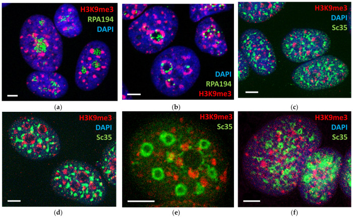
Figure 6.
Partial inhibition of rRNA and mRNA synthesis by AcD and alpha-amanitin revealed the hidden radial-concentric spatial relationship between centers of nucleolar synthesis, perinucleolar PADs, and speckles; this order was destroyed after full suppression of transcription: (a) Control: the granules of RPA1 fill the nucleoli, indicating active rRNA synthesis; (b) AcD 0.2 µM/mL, 1 h—RPA1 form rare fused granules, indicating suppressed rRNA synthesis; H3K9me3 PADs compact and encircle the nucleoli; (c) control: elongated speckles and PADs seem disorderly distributed in the cell nucleus; (d) AcD 0.2 µM/mL, 1 h—clumped speckles surround the compacted shells of perinucleolar PADs; (e) alpha-amanitin 2 µM/mL for 2 h suppressing Pol II—an example of swollen empty speckles radially circumventing the deteriorating perinucleolar ring of PADs; (f) AcD 2 µM/mL for 5 h suppressing both RNA syntheses with chaotically distributed disarranged PADs and empty speckles. Scale bars = 5 µm.

