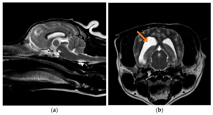
Figure 2.
(a) Midsagittal T2W MRI from a 7-year-old mixed-breed dog with Lafora disease; (b) transverse T2W MRI at the level of the temporal lobe from a 7-year-old mixed-breed dog with Lafora disease. There is dilatation of both lateral ventricles (orange arrow), which can be an early indicator of cortical atrophy associated with the disease. However, the size of the intrathalamic adhesion and subarachnoid space is normal.

