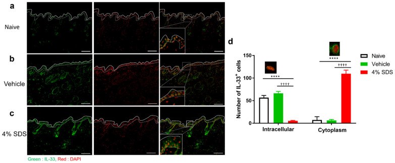
Figure 4.
Distribution of epidermal IL-33 in barrier-disrupted skin. (a–c) Immunohistochemical staining for IL-33 in skin lesions of naïve (a), vehicle-treated (b), and 4% SDS-treated (c) mice. Scale bars, 100 μm. (d) Epidermal IL-33 was measured and divided by the expression pattern of IL-33. n = 6–8. **** p < 0.0001 compared with naïve, †††† p < 0.0001 compared with vehicle-treated mice by two-way ANOVA with Tukey’s test. Numbers represent the mean ± SEM of two independent experiments.

