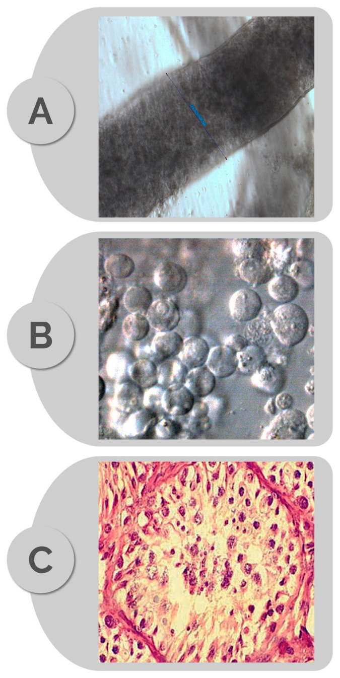
Figure 2.
Photomicrographs illustrating: (A) intact seminiferous tubule (diameter 270 micrometers), (B) cell suspension obtained after mechanical tubule mincing, and (C) corresponding histopathology (hematoxylin/eosin) specimen revealing germ cell maturation arrest (MA). Images A and B obtained at 400× magnification using an inverted optical microscope (Nikon Eclipse Diaphot 300, Nikon, Japan, with phase contrast (Hoffman)).

