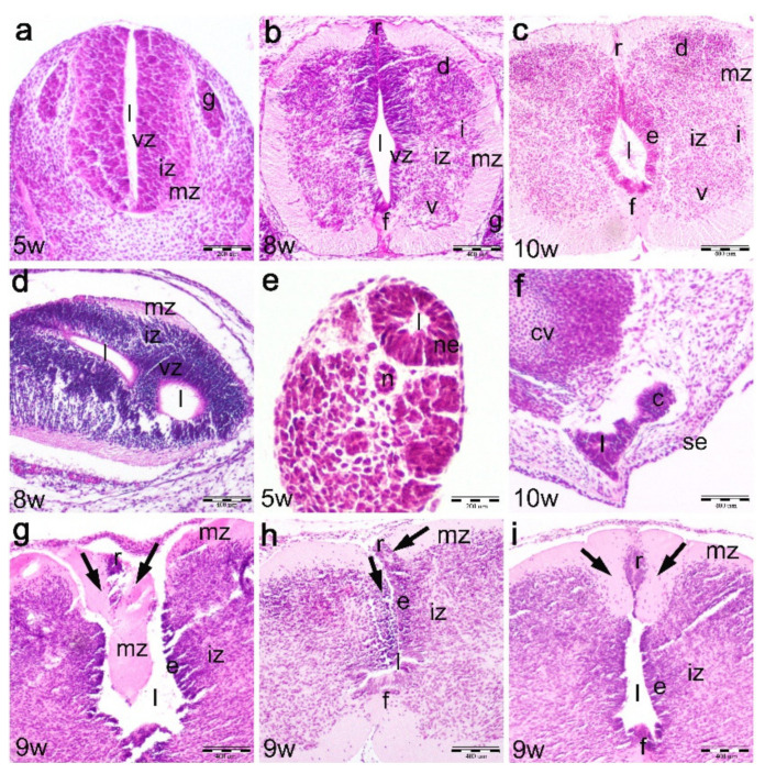
Figure 1.
Hematoxylin and eosin staining of the normal developing human spinal cord (5th–10th developmental weeks) and a 9th week human fetus with cervical spina bifida. Developing human cranial spinal cord (sc) in the 5th–6th weeks (a), 7th–8th weeks (b) and 9th–10th weeks (c), transitional zone in the 8th week (d) and caudal spinal cord in the 5th (e) and 10th week (f): ventricular zone (vz), intermediate zone (iz), marginal zone (mz), floor plate (f), roof plate (r), lumen (l), coccygeal remnant (c), coccygeal vertebrae (cv), skin epithelium (se), dorsal ganglia (g), ventral horns (v), intermediate horns (i) and dorsal horns (d), ependymal layer (e), notochord (n). Thoracic parts of the spinal cord in the 9th week fetus with cervical spina bifida (cranio-caudal direction) (g–i): ependymal layer (e), intermediate zone (iz), marginal zone (mz), roof plate (r), floor plate (f), lumen (l). Marginal zone abnormalities (arrows) in the roof plate area (r). Magnification ×20, scale bar 200 μm (a,e); magnification ×10, scale bar 400 μm (b–d,f–i).

