Figure 5.
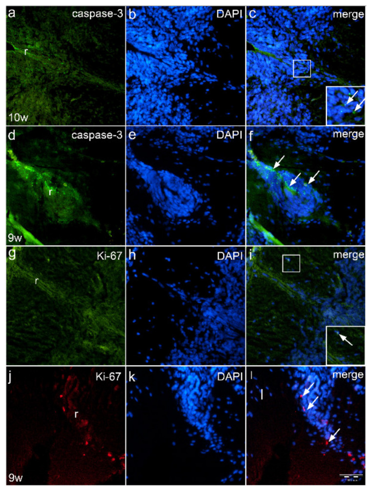
Immunofluorescence staining of roof plate area of normal human fetus and fetus with cervical spina bifida with apoptotic marker caspase-3 and proliferation marker Ki-67. Expression of caspase-3 apoptotic cells (arrows) in the roof plate (r) of normal 10th week embryo (a–c) has a different (regular) pattern when compared to the irregular distribution of caspase-3 positive cells (arrows) in the roof plate (r) of fetus with spina bifida (d–f). In normal fetus, Ki-67 positive cells are rarely seen (g–h), while in spina bifida, proliferating Ki-67 cells are irregularly organized (arrows) in the roof plate (r) (j–l). Magnification ×40, scale bar 100 μm (a–i).

