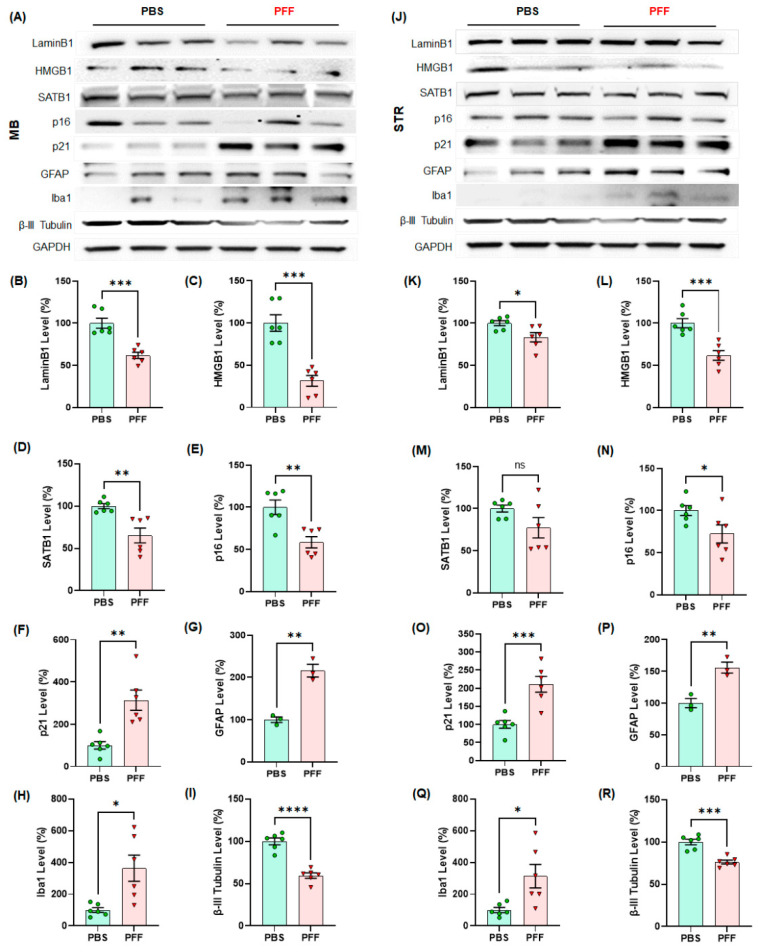
Figure 8.
The levels of cellular senescence markers such as LaminB1 and HMGB1 were significantly lower in α-syn PFF-injected mouse midbrain (MB) and striatum (STR) than PBS-treated brains, whereas the levels of p21, GFAP and Iba-1 were enhanced by 5–6 months after PFF treatment (n = 6/group). In Western blots, the examples of cellular senescence markers are displayed in triplicates per group ((A) MB, (J) STR). The quantified levels of Lamin B1 (B,K), HMGB1 (C,L), SATB1 (D,M), p16 (E,N), p21 (F,O), GFAP (G,P), Iba-1 (H,Q) and β-III-tubulin (I,R) are demonstrated. The level of β-III-tubulin decreased with α-syn PFF injection due to the neuronal loss. In quantification, the band intensity was normalized by a loading control, GAPDH and displayed in mean ± SEM in relativity (100% for age-matched controls, n = 6/group). The data analysis was applied to unpaired Student’s t-test for statistical significance. *: p < 0.05, **: p < 0.01, ***: p < 0.001 and ****: p < 0.0001. ns: not significant.

