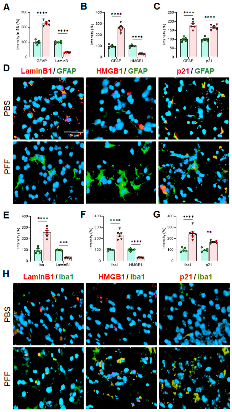
Figure 10.
In cell-type-specific analyses in the SNc, the levels of Lamin B1 and HMGB1 are significantly decreased in both astrocytes and microglia in α-syn PFF-injected mouse brains than PBS-treated ones, whereas the levels of p21 in both cell types are increased with α-syn PFF. In the SNc, both GFAP (A–C) and Iba-1 (E–G) label intensities were increased in PFF-injected SNc (n = 6/group) than PBS control. The intensities of Lamin B1 and HMGB1 in both astrocytes (A,B,D) and microglia (E,F,H) were reduced; however, the levels of p21 were significantly increased in both cell-types with α-syn PFF (C,D,G,H). DAPI stain (blue) was used to indicate the location of nucleus. The label intensities of Lamin B1, HMGB1 and p21 were cell-type specifically quantified, based on double-labels in a blinded manner. In quantification, PBS injected SNc region was used as a relative label intensity (100%, n = 5/group) in mean ± SEM and applied to unpaired Student’s t-test for statistical significance. **: p < 0.01, ***: p < 0.001 and ****: p < 0.0001. Size bar: 100 µm.

