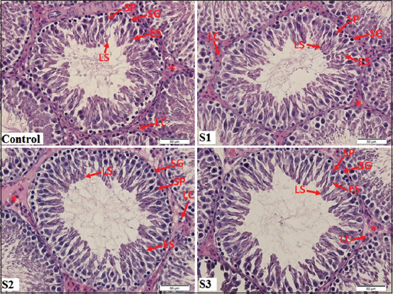
Figure-2.

Seminiferous tubule section in rats. (H & E staining [400× magnification]). H & E staining showing the seminiferous tubules and the interstitial tissue (asterisks) containing the Leydig cells (LC). Each tubule is lined with Sertoli cells (SC), spermatogonia (SG), primary spermatocytes (SP), early spermatids (ES), and late spermatids (LS). The seminiferous tubules of control group showed germ cells organized in concentric layers and tubular lumen was empty. The administration of only Marsilea crenata in healthy rat was not significantly different with control group in the spermatogenesis condition.
