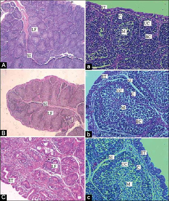
Figure-9.

Histological structure of BF of a control chicken at three different ages (14, 28, and 42) with two magnifications. (A, B, C 4×) and (a, b, c 20×). SI: Space interfollicular, C: Cortex, M: Medulla, LF: Lymphoid follicle, ET: Epithelial tissues, UC: Undifferentiated cells, BC: Blood capillaries; BF: Bursa of fabricius.
