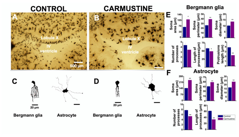
Figure 3.
Carmustine treatment reduced morphological complexity of glial cells. (A). Coronal section at the level of lobule X of the cerebellum processed by Golgi-Cox staining. (B). A sample image of the same region showing a cortical dysplasia. Notice the malformation of the shape of the fourth ventricle. (C,D). Representative drawings with camera lucida of a Bergmann cell of lobule X and an astrocyte from cerebellar cortex, control and carmustine respectively. Complexity and extension of the astrocyte processes are observed in the control group, which contrasts with the carmustine-treated group, as fewer and diffuse processes are observed, as well as an increase in the size of the soma. (E,F). Comparative analysis between the control and the carmustine-treated groups (1 to 3 BG cells were analyzed from each experiment; Control n = 6, Carmustine n = 6 and 6 to 8 astrocyte cells were analyzed from each experiment; Control n = 6, Carmustine n = 6). Data are represented as the mean ± the standard error of the mean. * indicates significant difference (p < 0.05) and ** indicates significant difference (p < 0.001) both compared to the control group.

