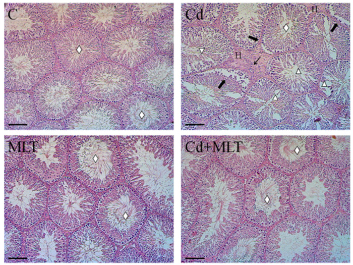
Figure 1.
Hematoxylin-eosin staining of C-, Cd-, and/or MLT-treated rat testis. Evaluation of testicular histology of animals exposed to Cd and/or MLT. Rhombus: tubules lumen; Thick arrow: GC desquamation; Triangle: space between GC; Thin arrow: mononuclear cell infiltration; H: hemorrhage. Scale bars represent 40 µm.

