Figure 3.
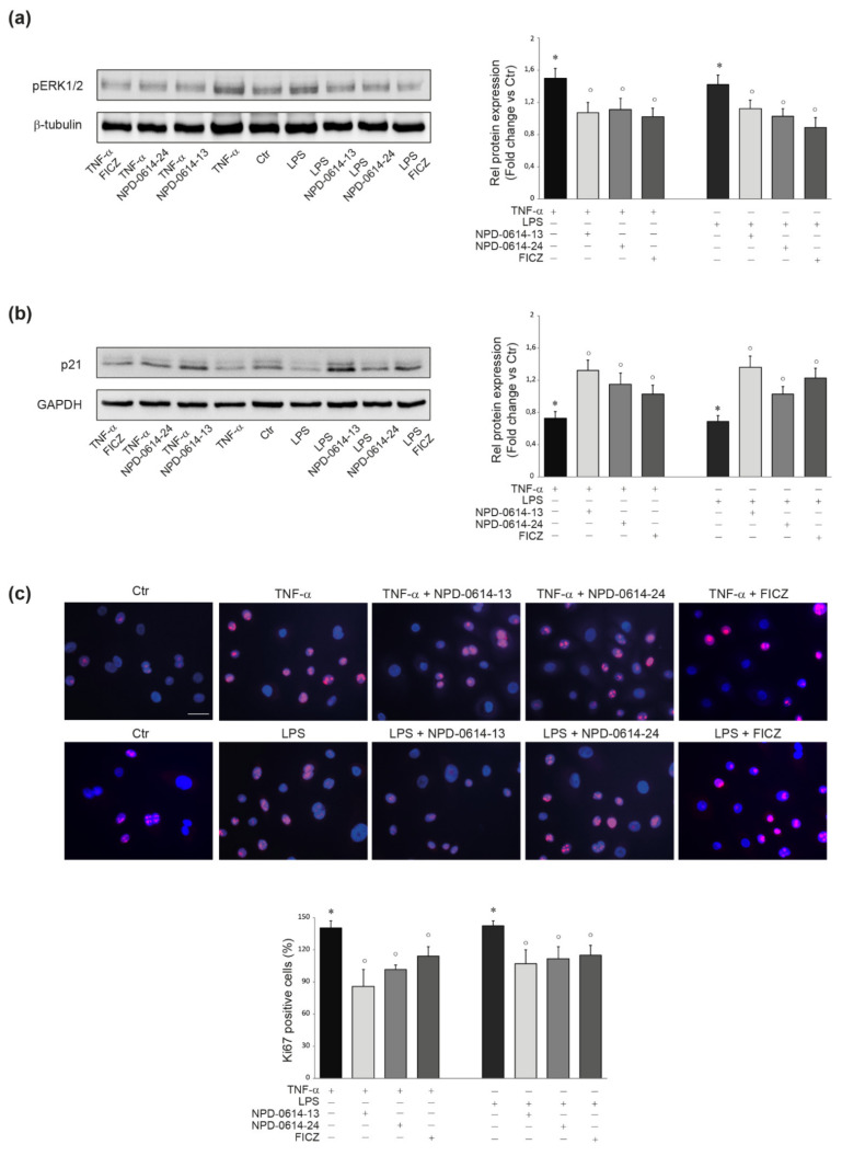
NPD-0614-13 and NPD-0614-24 recovered the altered proliferation induced by TNF-α or LPS in NHKs. (a) Western blot analysis and corresponding densitometric analysis of pERK1/2 protein expression in NHKs treated with TNF-α (20 ng/mL) or LPS (10 μg/mL) in the presence or absence of NPD-0614-13, NPD-0614-24 (25 μM) and FICZ (100 nM) for 1 h. (b) Western blot analysis and corresponding densitometric analysis of p21 protein expression in NHKs treated as above for 6 h. β-tubulin or GAPDH were used as endogenous loading control. Results are expressed as the fold change respect to untreated control cells. Data represent the mean ± SD of three independent experiments (significance vs. untreated control or vs. stimulated cells are marked with * and °, respectively; * p < 0.05 vs. untreated cells; ° p < 0.05 vs. TNF-α or LPS stimulated cells). (c) Representative immunofluorescence images and fluorescence quantitative analysis of Ki67 expression in NHKs treated as above for 48 h. Nuclei were counterstained with DAPI. Bar: 20 μm. Results are expressed as the mean percentage of Ki67-positive cells (* p < 0.05 vs. untreated cells; ° p < 0.05 vs. TNF-α or LPS stimulated cells).

