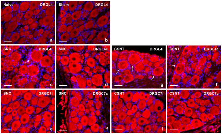
Figure 1.
Representative sections of DRG from naive rat (a) and rats with sterile sham- (b), SNC- (c–f) or CSNT- (g–j) interventions for 7 days. The DRG sections of lumbar (DRG-L4) and cervical (DRG-C7) segments of both ipsilateral (i) and contralateral (c) sides were immunostained for TLR9 using a rabbit polyclonal antibody and donkey TRITC-conjugated anti-rabbit secondary antibody under the same conditions. Besides neuronal bodies, increased TLR9 immunofluorescence was found in satellite glial cells enveloping the neuronal bodies of ipsilateral DRG-L4 after SNC (arrow in Figure 1c) and bilaterally in DRG-L4 after CSNT (arrows in Figure 1g,h). Scale bars = 40 µm.

