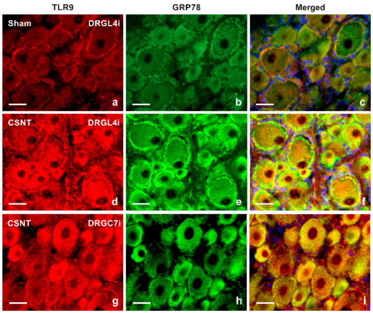
Figure 3.
Double immunofluorescence staining for TLR9 and GRP78 as a marker of the endoplasmic reticulum (ER). Sections of lumbar DRG ipsilateral (DRGL4i) to sham operation (Sham) as well as DRGL4i and cervical DRG ipsilateral (DRGC7i) to CSNT were immunostained with mouse monoclonal anti-TLR9 (a,d,g) and rabbit polyclonal anti-GRP78 (b,e,h) antibodies and were visualized using donkey TRITC-conjugated anti-mouse and FITC-conjugated anti-rabbit secondary antibodies, respectively. Cell nuclei were stained with Hoechst 33342. Merged pictures (c,f,i) detected no or very weak TLR9 immunostaining in the ER of DRG neurons from the sham-operated animal (c), while most increased TLR9 immunofluorescence was co-localized with GRP89 in the ER of DRGL4i (f) and DRGC7i (i) from CSNT-operated rats. Scale bars = 20 µm.

