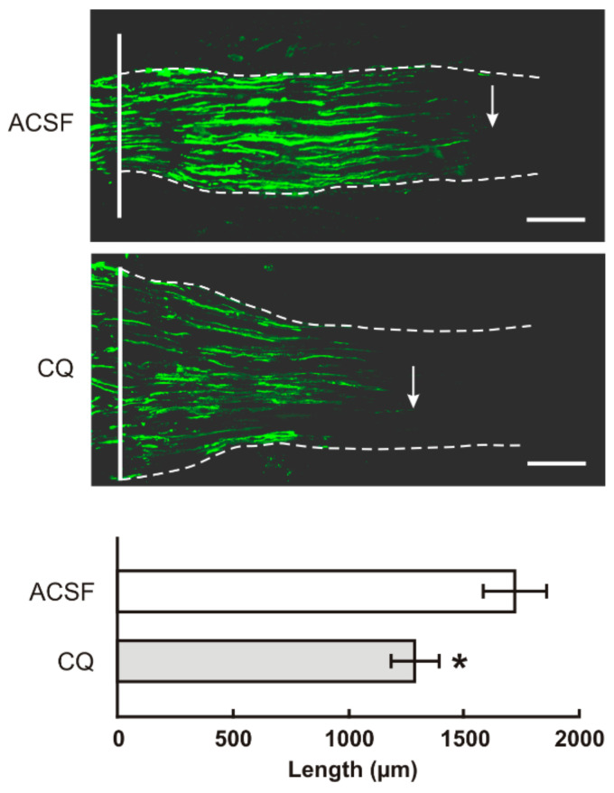
Figure 7.
The top panels illustrate representative longitudinal sections through ulnar nerves distal to the crush site (solid line) showing SCG10 immunopositive regenerated axons. Arrows indicate the tip of the longest SCG10 immunopositive axons regenerated 1 day after the ulnar nerve crush in rats with prior CSNT for 7 days and intrathecal application of artificial cerebrospinal fluid (ACSF) or chloroquine (CQ). Scale bars = 280 µm. The bottom panel illustrates mean length of regenerated SCG10 immunopositive axons ± SD in the ulnar nerve 1 day after crush and 7 days from intrathecal application of artificial cerebrospinal fluid (ACSF) or CQ; n = 6 for each group. * Significant difference (p < 0.05) compared with ACSF in a Kruskal–Wallis ANOVA test.

