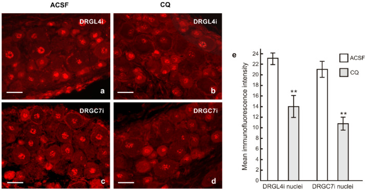
Figure 9.
Representative sections of the lumbar (DRGL4i) and cervical DRG (DRGC7i) ipsilateral to the crushed ulnar nerve in rat with prior CSNT for 7 days and intrathecal application of artificial cerebrospinal fluid (ACSF, (a,c)) or chloroquine (CQ, (b,d)). Sections were immunostained with rabbit polyclonal anti-STAT3(Y705) and TRITC-conjugated donkey anti-rabbit antibodies under the same conditions to demonstrate nuclear translocation of activated STAT3. Scale bars = 40 µm. The graph (e) shows decreased intensity of STAT3 immunofluorescence in the DRG neuronal nuclei after CQ application. ** Significant difference (p < 0.01) compared with ACSF in a Kruskal–Wallis ANOVA test.

