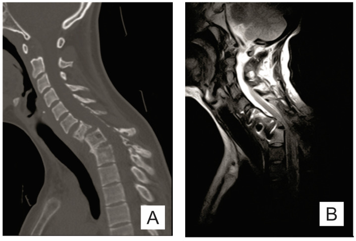
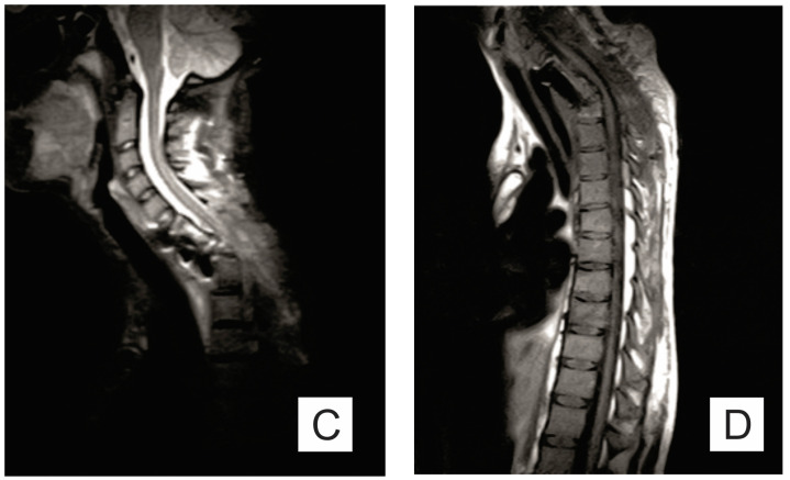
Figure 1.
(A) Sagittal CT scan: osteolytic lesions (C6–C7, T1–T2), pathological fracture of the C7 vertebra; (B) sagittal T2 MRI; (C) sagittal STIR MRI; and (D) sagittal T1 MRI: important spinal canal stenosis at C6–C7 level up to an anteroposterior diameter of 5 mm, the medullary cord presents in this area discreetly T2-STIR hypersignal.


