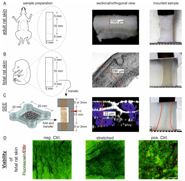
Figure 3.
Skin model systems to analyze dermal and epidermal mechanical properties. Three models of different maturation state and composition were comparably prepared: (A) Adult rat skin explants were excised from back skin and the test dimension used for analysis previously marked in situ. The mean dermo-epidermal thickness (exemplarily labeled with red line) was manually determined from tissue slices (n = 16). Underlying hypodermal tissue was omitted here; (B) Back skin from embryonic day 18 rats was cut into indicated test dimensions after isolation. Analysis length was set to 10 mm initial length using a C-shaped PVDF frame (gray). Mean tissue thickness was measured from 14 independent sections (n = 14); (C) Similar PVDF frames were used for transfer of multilayered, simplified epidermis equivalents (SEEs). Therefore, the 20 × 20 mm cell sheets were folded 2 times along one axis to obtain the 5 × 20 mm test dimension. The mean cell sheet thickness is indicated (n = 17); (A–C) Right images show mounted samples previous to start of the test protocol. The lateral white PVDF transfer frame in fetal skin and SEE samples were cut after mounting and had therefore no influence on subsequent force measurements. Scale: 2 mm. Red label illustrates the outline of SEE; (D) Viability assay of epidermal cells via immunofluorescence was performed with fetal rat skin after transfer and stretching for at least 4 h. Non-transferred but identically prepared samples served as controls. Fetal skin in positive controls were deliberately killed with azide. Scale: 50 µm.

