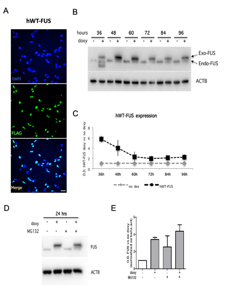
Figure 1.
Human WT-FUS is expressed in mouse NPSCs. (A) Immunostaining using an anti-FLAG antibody on proliferating NSPCs electroporated with the hWT-FUS plasmid, showing the nuclear localization of the exogenous protein. Cell nuclei are stained with the nuclear dye Hoechst. Scale bar 30 µm. (B,C) Representative immunoblot of proliferating NSPCs in time course between 36 and 96 h from transgene induction (B) and densitometric analysis of hWT-FUS transgene expression (C). (D,E) Representative immunoblot of proliferating NSPCs grown for 48 h and then treated with MG132 (0.2 uM) for 24 h and densitometric analysis of FUS levels. Beta-actin is used as a loading control. Values are expressed as the average of three independent experiments. Data are presented as a mean ± SEM.

