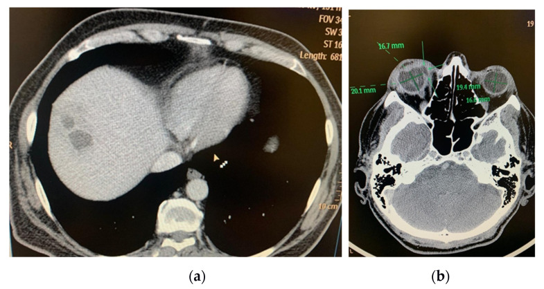
Figure 1.
(a) CT exam: multiloculated hepatic liver abscess, right lobe, segment VIII, 50/32/57 mm; (b) CT Exam: Right eye endophthalmitis, eyeball with irregular contour, with a hypodense area in the internal side (at the area of the insertion of the medial rectus muscle), with possible communication between posterior chamber and periorbital space (perforation) (archive of the Ophthalmology Department, Emergency University Hospital Bucharest).

