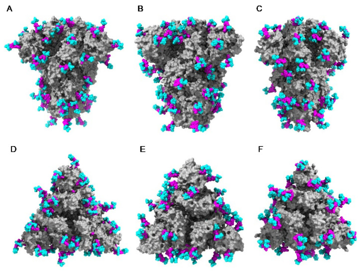
Figure 35.
(A–C). Lateral views of the molecular surfaces (colored grey) of spikes from SARS-CoV (PDB code 6ACD) (A), MERS-CoV (PDB code 5W9H) (B), and SARS-CoV-2 (PDB code 6VXX) (C), showing the exposure of the fucosylated and non-fucosylated tri-mannosyl cores (colored magenta) of high mannose glycans and complex glycans (colored cyan). (D–F). Front views of the molecular surfaces (colored grey) from SARS-CoV (D), MERS-CoV (E), and SARS-CoV-2 (F), showing the exposure of the fucosylated or non-fucosylated tri-mannosyl cores (colored magenta) of high mannose glycans and complex glycans (colored cyan).

