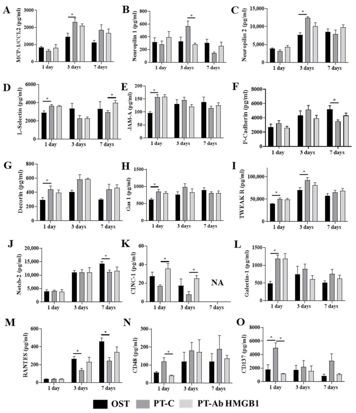
Figure 2.
Plasma samples from osteotomy (OST), polytrauma (PT-C), and polytrauma + anti-HMGB1 antibody (PT-Ab HMGB1) at 1, 3 and 7 days post-trauma (dpt) were quantitatively assessed for cytokine protein expression. (A) Monocyte attracting chemokine (MCP-1/CCL2); (B) Neuropilin 1; (C) Neuropilin 2; (D–F) Adhesion molecules: L-selectin, junctional adhesion molecule (JAM-A) and P-cadherin, respectively; (G) Proteoglycan: decorin; (H) Cell arrest and apoptosis regulator (GAS-1); (I) TNF-related weak inducer of apoptosis receptor (TWEAK R); (J) Neurogenic locus notch homolog protein 2 (Notch 2); (K) Cytokine-induced neutrophil chemoattractant 1 (CINC-1); (L) Anti-inflammatory and T cell suppressive protein: Galectin 1; (M) Regulated on activation, normal T cell expressed and secreted (RANTES); (N) Lymphocyte activation marker: CD48; and (O) lymphocyte activation co-stimulatory immune checkpoint molecule: CD137. (n = 4–5 for OST, PT and PT-Ab HMGB1) * p < 0.05 comparing OST and PT-Ab HMGB1 rats to PT-C rats. The bar graphs represent the mean, whereas error bars represent SEM. NA–protein expression data is not available.

