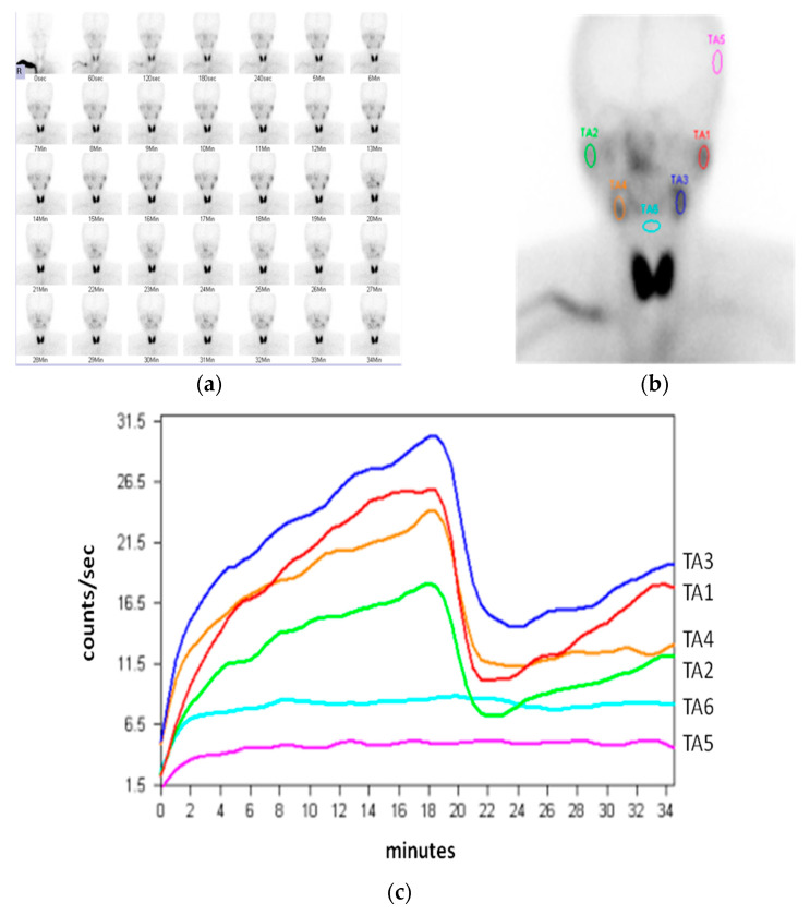
Figure 1.
(a) Sequential imaging in sialoscintigraphy; (b) region of interest (ROI) positioned at the salivary glands, left temporal region of skull, and hypopharynx, respectively; TA1: left parotid; TA2: right parotid; TA3: left submandibular; TA4: right submandibular; TA5: temporal region as the background of the parotid gland; TA6: hypopharynx as the background of the submandibular gland; (c) time–activity curves (TACs) generated from six ROIs.

