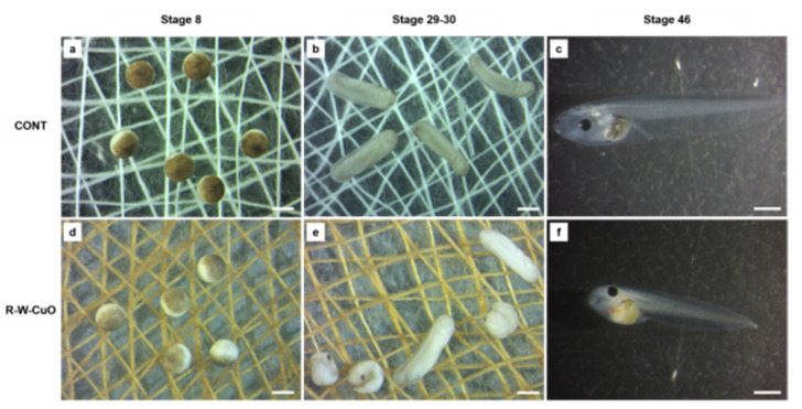
Figure 6.
Stereomicroscopic images showing the experimental set up, with embryos in direct contact with uncoated cotton bandages (a,b) and CuO NP-coated bandages in water (W-CuO) (d,e), fixed at the bottom of glass petri dishes. Embryos were photographed at the beginning (stage 8) (a,d) and at the middle (stage 29–30 of the test). Larvae at the end of the test (stage 46) were screened for single malformations: (c), control larva; (f), W-CuO exposed larva showing irregular gut and body length shortening. Bars = 1 mm.

