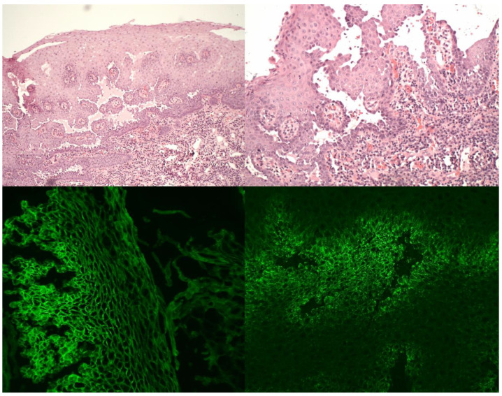
Figure 1.
Histopathology and direct immunofluorescence of laryngeal pemphigus vulgaris specimens. Upper left—suprabasilar acantholytic blister with intraepithelial suprabasilar clefting; HE stain, 100× magnification. Upper right—suprabasilar acantholysis and plasma cell infiltrate among villous-like projections of lamina propria, covered by a few layers of epithelium; HE stain, 200× magnification. Lower left—intercellular linear deposition of IgG in direct immunofluorescence, DIF IgG, 200× magnification. Lower right—intercellular granular deposition of C3 in direct immunofluorescence, DIF C3, 200× magnification.

