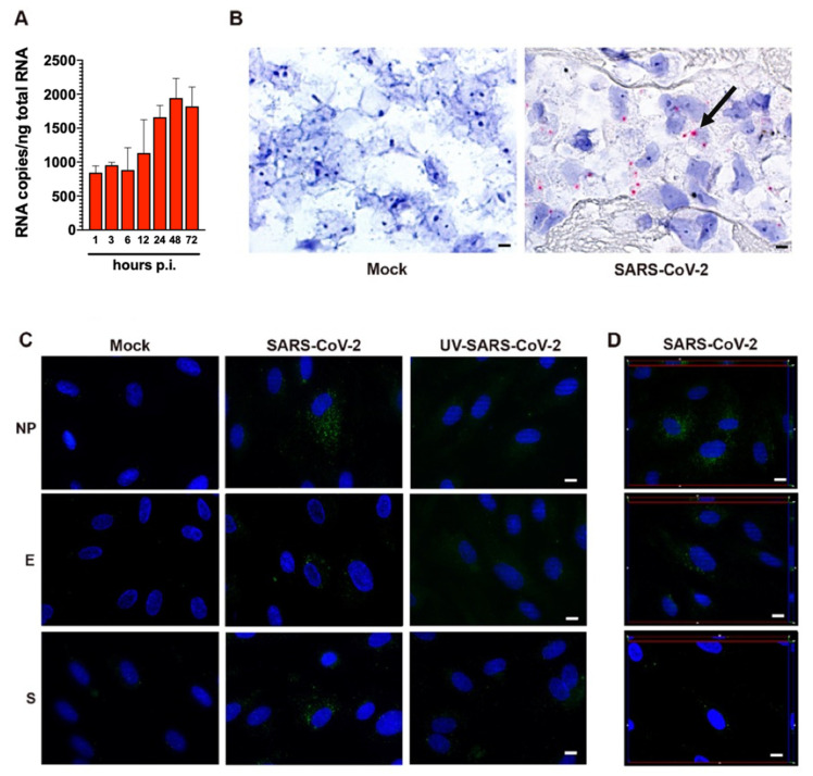
Figure 2.
SARS-CoV-2-infected HL-mECs express viral RNA and proteins. (A) Quantitation of SARS-CoV-2 genomes at the intracellular level by qRT-PCR. At least three replicates were performed. Data are representative of two independent experiments with similar results. (B) SARS-CoV-2 RNA in situ hybridization using the S antisense probe shows strong expression of viral RNA as red cytoplasmic dots in SARS-CoV-2-infected HL-mECs (black arrow), while mock-infected cells (Mock) are negative (scale bar, 3 µm). (C) HL-mECs were mock-infected (Mock) or infected with SARS-CoV-2 (SARS-CoV-2) or with UV-inactivated SARS-CoV-2 (UV-SARS-CoV-2) at MOI 1, for 1 h at 37 °C, then washed and cultured until day 3 p.i. Immunofluorescence was performed by incubating cells with antibodies targeting the SARS-CoV-2 NP (upper panels), E (middle panels), or S (lower panels) proteins (scale bar, 10 µm). (D) z-Stack sections and orthogonal z reconstitution of SARS-CoV-2-infected HL-mECs expressing NP, E, or S proteins (scale bar, 10 µm). Images of HL-mECs display SARS-CoV-2 signals in green and cell nuclei in blue.

