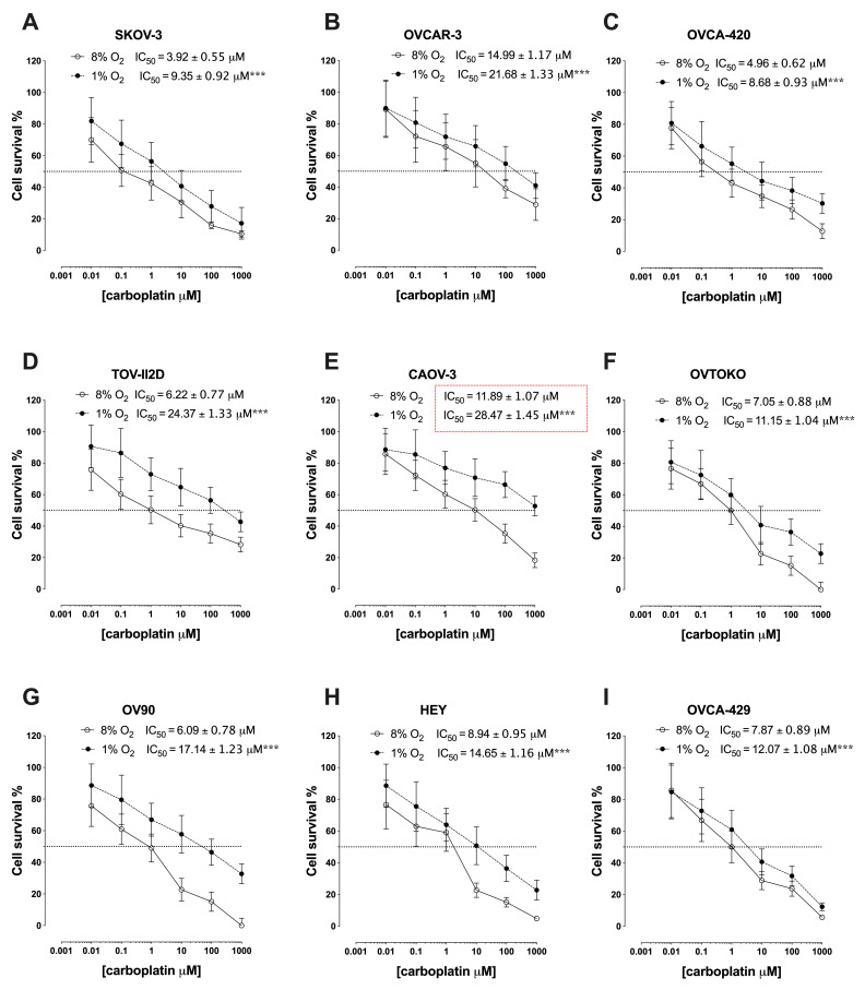
Figure 1.
IC50 of a panel of epithelial ovarian cancer cells. The cells ((A–I): SKOV-3, OVCAR-3, OVCA-420, TOV-112D, CAOV-3, OVTOKO, OV-90, HEY, and OVCA-429) were exposed to either 1% O2 or 8% O2 and treated with carboplatin at different concentrations (0, 0.001 µM, 0.01 µM, 0.1 µM, 1 µM, 10 µM, 100 µM, and 1000 µM) in the presence of Caspase-3/7 reagent. The viability of the cells was assessed using the IncuCyte™ real-time cell-imaging system every 2 h for 72 h. The data are represented by the mean ± SEM (n = 6). The IC50 of all EOC cells in this study were higher under hypoxia than under normoxic in their parental cells. *** p < 0.0005 at 1% oxygen compared with 8% oxygen.

