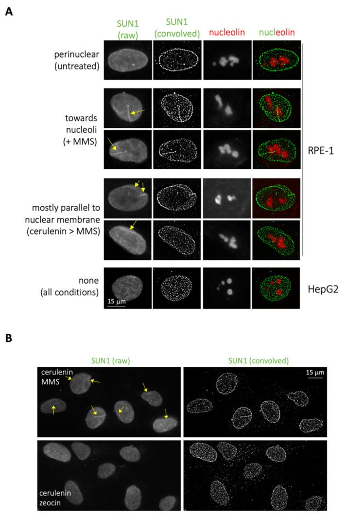
Figure 3.
Increase in intranuclear membrane pools occurs in RPE-1 cells in response to MMS. (A) Immunofluorescence performed on RPE-1 and HepG2 cells to detect SUN1 and nucleolin signals. Visualization of membrane-associated SUN1 signals is enhanced by convolving the raw image. In RPE-1 cells, in untreated conditions, SUN1 signals are mostly perinuclear; in response to MMS, SUN1 invaginations reach nucleoli; preincubating the cells with cerulenin prior to MMS addition, SUN1 invaginations mainly run parallel to the nuclear membrane. In HepG2 cells, SUN1 antibody provided no membrane-associated pattern under these immunofluorescence conditions. (B) Immunofluorescence to detect SUN1 performed on RPE-1 cells treated 4 h with 5 μg/mL, the last 2 h either with additional 0.005% MMS or 100 μg/mL zeocin. On the left, raw images are shown, and NR signals are indicated by yellow arrows. On the right, the same images were subjected to the ImageJ tool “convolve”.

