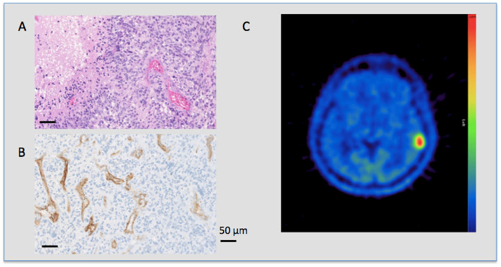
Figure 3.
PSMA expression and metabolic tumor activity in a glioblastoma patient. HE (upper left side) and PSMA (lower left side) staining and the corresponding MET PET scan (right side) of a 58 year old patient with an IDH-R132H negative GBM. The patient died 19 months after diagnosis. (A) HE staining in 20-fold magnification of a GBM showing a typical crowded, polymorphic cell count and dense vessels. (B) PSMA IHC also in 20-fold magnification of the same patient showing an intense reaction on the vascular endothelium with no staining of non-vascular cells. (C) MET PET scan of the same patient showing a high focal uptake in the tumor region in the left temporal lobe with a high T/N ratio value of 4.98.

