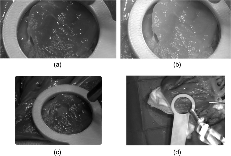
Fig. 4.
Spatially resolved HMSI images of the heart of the third pig model. (a) Multispectral VIS camera; (b) multispectral NIR camera; and (c) Multispectral VIS camera. One spectral band of the snapshot sensor data for (a) , (b) , and (c) . (d) The intensity distribution of the TIVITA Tissue pushbroom camera image at .

