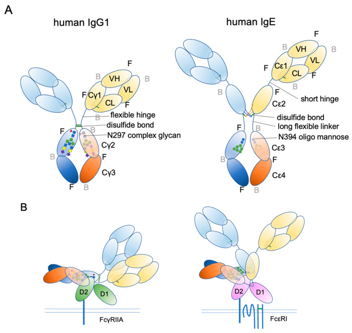
Figure 3.
Schematic representation of human IgG1 and IgE. (A) The extended conformations. The three-dimensional coordination (front or back) is shown. F, front; B, back. (B) The receptor bound conformations. Please note that IgE Fab is almost in the upright position, suitable for sensing its target antigens.

