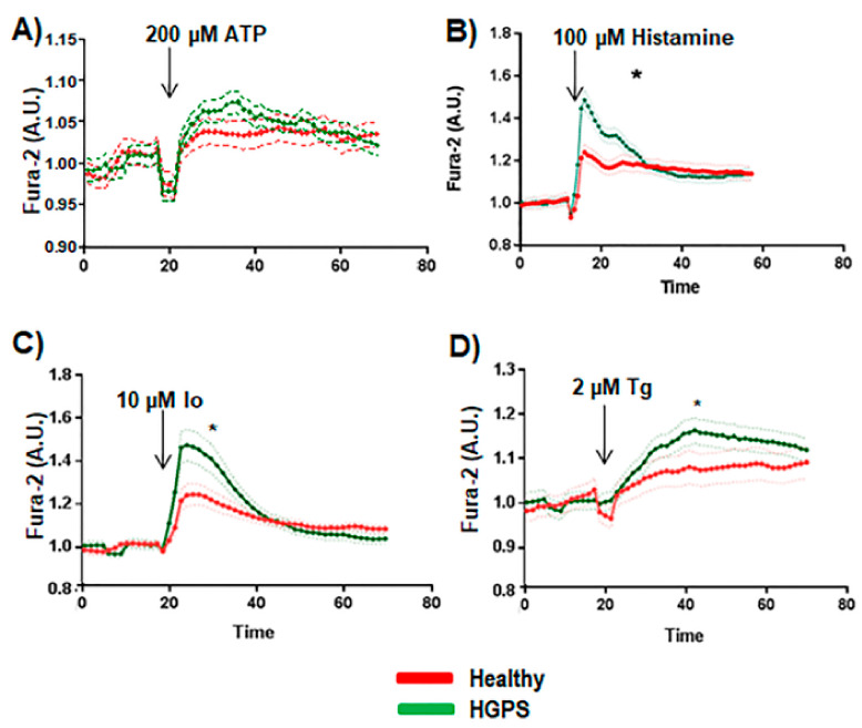
Figure 3.
Cytosolic calcium handling study. (A) Ca2+ flux in the absence of extracellular calcium with 3 mM of EGTA in the medium was performed using healthy (AG03257, AG03258, AG03512, and AG06299) and HGPS (AG03198, AG03199, AG03513, and AG06917) human skin fibroblasts in a FlexStation reader. (B) Results after adding ATP at 100 μM (arrow). (C) Results after adding 10 μM ionomycin (Io) (arrow). (D) Results after adding 10 μM thapsigargin (Tg) (arrow). The average calcium baseline levels were 33.61 ± 6.5 nM for WT cells and 76.21 ± 9.09 nM for HGPS cells. Bars are means ± SEM from three independent experiments. * p < 0.001 was considered statistically significant.

