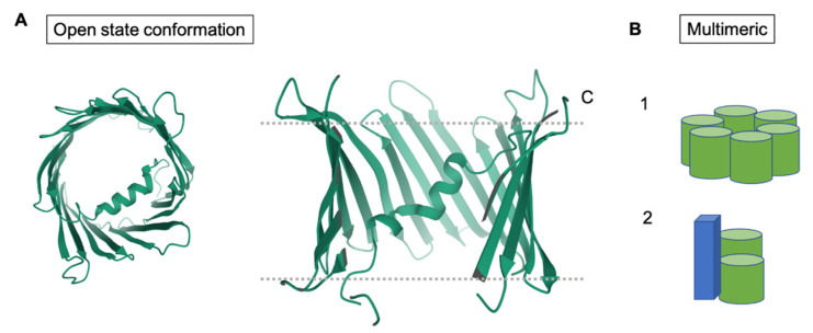
Figure 5.
Structural conformations of VDAC. (A) Shows top and side views of hVDAC1 in the open conformation (PDB ID: 2JK4). (B) Models of the oligomeric structures of VDAC, including the homomeric and hexameric forms. The green cylinders represent VDAC. The blue cuboid represents a protein, other than VDAC, that associates to form a hexameric complex.

