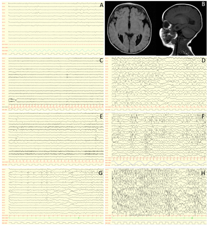
Figure 2.
EEG evolution at 0 (A), 3 (C,D), 5 (E,F) and 9 (G,H) years old, and RM features (B) of a MWS individual. Caption: At birth, electroencephalographic activity is essentially normal (A). With time, a worsening is observed, with an increase in paroxysmal abnormalities in wakefulness (C,E,G), dramatically activated during sleep (D,F,H). MRI images performed at 4 months show complete agenesis of the corpus callosum (B). Interestingly, abnormalities in sleep can sometimes appear asynchronous although bilateral: this is probably related to this type of malformation. Note: low frequency filter: 1.6 Hz; high frequency filter: 60 Hz; paper speed: 20mm/sec; sensitivity: 150 microV/cm (A,C,D,E,F), 200 microV/cm (G) and 100 microV/cm (H).

