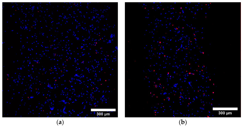
Figure 7.
Live/dead fluorescence images (10× magnification) of (a) 3/4 Alg/Gel and (b) 3/7 Alg/Gel bioinks measured immediately after extrusion. Blue color represents all cells, alive and dead, stained with Hoechst 33342, and red color represents propidium-iodide-stained dead cells. The scale in both figures is 300 µm.

