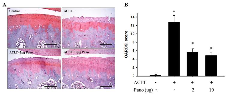
Figure 3.
Histopathological evaluation of the tibia in knee joints after panobinostat treatment in an ACLT rat model. (A) Safranin O/Fast Green staining was performed on the histological sections of knee joints from the control, ACLT, and ACLT + panobinostat (2 or 10 μg) groups. Representative images of Safranin O/Fast Green staining for articular cartilage show cartilage damages in the ACLT knee compared with panobinostat treatment. The scale bar represents 250 μm. (B) Histopathological changes in the knee joints of the four studied groups were evaluated using the OARSI scoring system. Histogram shows the OARSI scores of the control, ACLT, and ACLT + panobinostat (2 or 10 μg) groups. OARSI, Osteoarthritis Research Society International; ACLT, anterior cruciate ligament transection; Pano, panobinostat. (* p < 0.05 vs. the control group; # p < 0.05 vs. the ACLT group).

