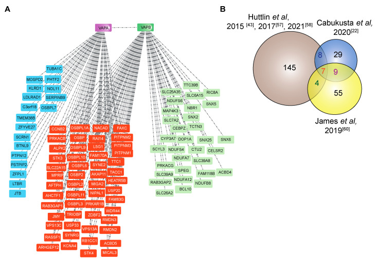
Figure 4.
The interactome of VAPA and VAPB. (A) The Bioplex interaction network of both VAPA and VAPB in HEK293T cells reported by Huttlin et al. [43,57,58]. The proteins represented in blue are sole VAPA interactors, and those represented in green are sole VAPB interactors. The proteins represented in orange are common interacting partners of both VAPA and VAPB. (B) The Venn diagram shows VAPB interactors identified by Huttlin et al. (affinity purification-mass spectrometry) [43,57,58], by Cabukusta et al. (BioID followed by mass spectrometry identification) [22] and by James et al. (RAPIDS (rapamycin and apex dependent identification of proteins by SILAC) followed by mass spectrometry identification) [60]. See Table 1 for proteins that were identified in at least two studies.

