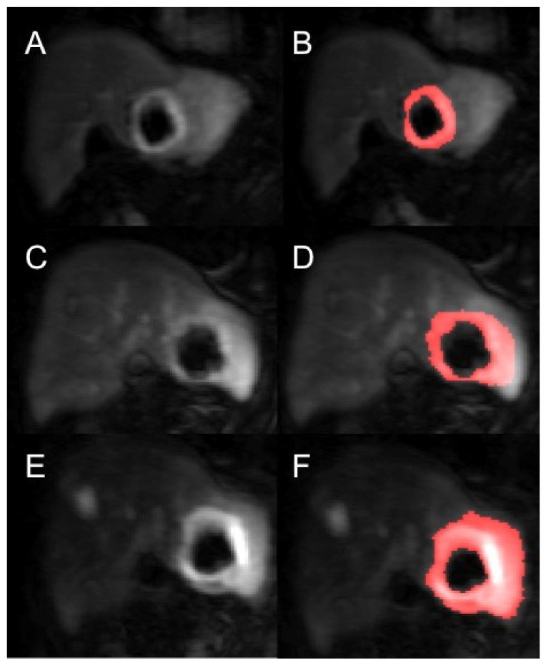
Figure 2.
Example of a segmented renal metastasis on DCE-MRI within the left lobe of liver (before and after segmentation with thresholding technique of tumour ROI—the red highlighted area is the tumour ROI segmented on the PMI software) at baseline (A,B), 4-weeks (C,D) and 10-weeks (E,F). This is a case of disease progression at 6 months.

