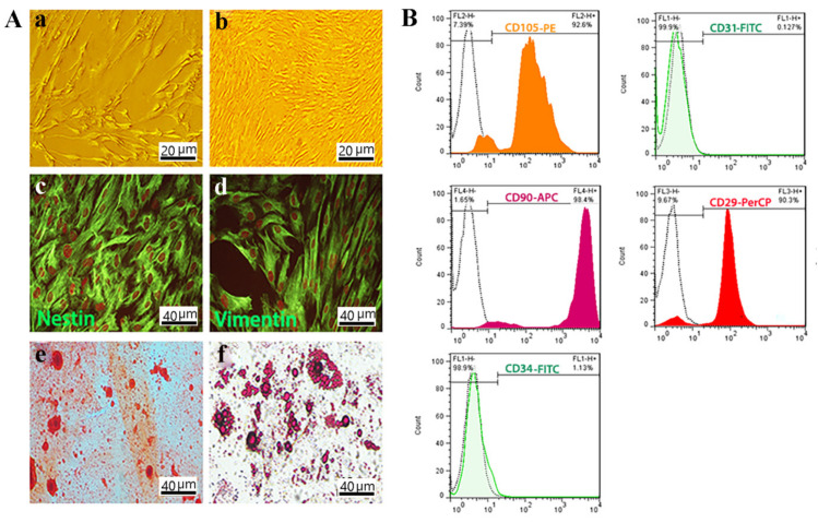
Figure 2.
OE-MSCs characterization. (A) Morphology of OE-MSCs in passage one. Scale bar: 20 μm; (a,b) Morphology of OE-MSCs in passage three which was described as a spindle-like morphology. Scale bar: 10 μm; Immunocytochemistry of OE-MSCs to characterized specific neural crest and mesenchymal markers; (c) nestin; and (d) vimentin by immunofluorescence staining. Osteogenic and adipogenic differentiation of OE-MSCs, respectively; (e,f) Scale bar: 40 μm. (B) Flow cytometric assessment to characterization of OE-MSCs surface markers; including CD29 as a positive marker; CD31 as a negative marker; CD105 as a positive marker; CD90 as a positive marker; and CD34 as a negative marker.

