Figure 2.
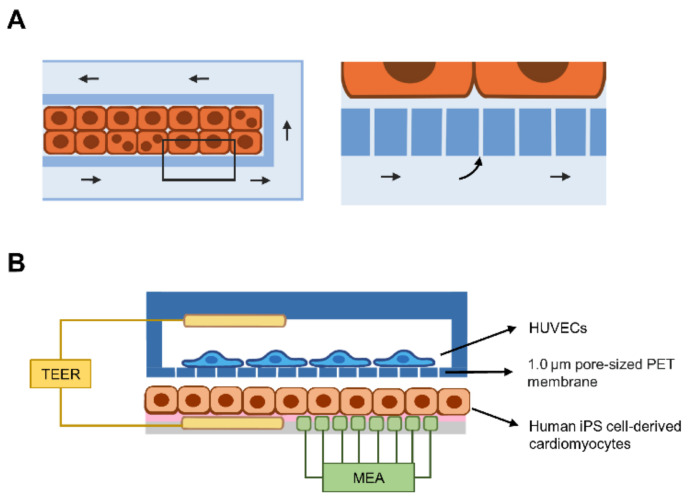
Current liver-on-a-chip models for hepatotropic infectious diseases and a combination of biosensors and organs-on-chip. (A) The microfluidic chip contains two compartments: a microchamber for hepatocyte culture (orange-colored rectangles) and microchannel (light blue) for fluid flow (arrows). The magnified box shows the porous layer that recapitulates the fenestration of SECs and allows the diffusion of culture medium into the cell-filled microchamber. (B) Coculture of human iPS cell-derived cardiomyocytes and HUVECs in double microchambers. Human primary hepatocytes (orange-colored rectangles) are grown on the fibronectin-coated surface (pink) of the lower chamber, while HUVECs are grown on the porous PET membrane of the upper chamber. The transepithelial electrical resistance (TEER, yellow) electrode is inserted into the upper and lower chambers. Gap formation between endothelial cells causes a decrease in TEER. Moreover, a multielectrode array (MEA) can be inserted in the lower chamber to detect the beating rate of cardiomyocytes. Abbreviations: HUVEC, human umbilical vascular endothelial cell; PET, polyethylene terephthalate.

