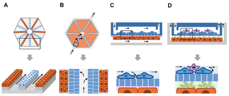
Figure 4.
Design of a liver-on-a-chip based on the microarchitecture of the liver lobule. (A) The radial pattern of hepatocytes and SECs (light blue-colored ovals) is modeled using electric-based cell seeding. The magnified box shows two lines of hepatocytes and a line of endothelial cells. (B) Hexagon-shaped microchip containing six microchambers (red) for cell seeding. The magnified box shows a double porous layer (blue-colored rectangles with white lines) as the fenestrated SECs. Due to their distal and proximal locations to the porous layer of hepatocytes, nutrients and gas diffuse into hepatocytes in a gradient pattern (arrows). (C) The two-sided layer allows coculture of distinct cell types. Human primary hepatocytes (orange-colored rectangles) are grown on collagen-coated surfaces (pink), while immortalized bovine aortic endothelial cells are grown in the upper chamber. A porous PET membrane separates the two chambers and allows small molecules to pass through. (D) Heterogeneous cell culture in a double microchamber. Human primary hepatocytes (orange-colored rectangles) are grown on the fibronectin-coated surface (pink) of the lower chamber, while the endothelial cell line is grown on the porous PET membrane of the upper chamber. The space between hepatocytes and the porous upper layer is filled with collagen gel (light green) containing hepatic stellate cells (reticular, green-colored shape). The human monocytic cell line U-937 (violet-reticular shapes) was cultured on top of ECs to mimic liver-resident macrophages (Kupffer cells). Arrows indicate the direction of fluid flow.

