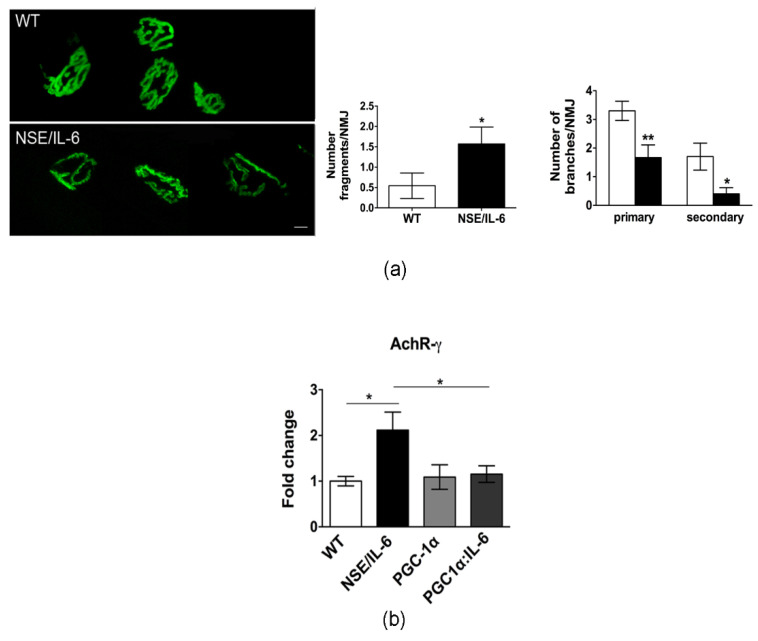
Figure 5.
IL-6 overexpression affects neuromuscular junction (NMJ) destabilization. (a) Confocal microscopy of post-synaptic NMJ stained with a-bungarotoxin; representative images of one spatial series (from 0 to 50 µm) composed of 25 optical sections with a step size of 2 µm, from tibialis anterior of 6-month-old wild type and NSE/IL-6 mice. Scale bar 10 μm. Graphs showing the degree of NMJ fragmentation (Number fragments/NMJ) and the topology of branching pattern (Number of branches/NMJ). Data are represented as average ± SEM n = 3; * p< 0.05, ** p < 0.01. Statistical significance assessed by Mann–Whitney Rank Sum Test. (b) Real time PCR for the expression of AchR-γ in soleus wild type (WT) and NSE/IL-6, PGC-1α, and PGC1α:IL-6 mice at 6 months of age. Values are expressed as fold change variations relative to WT and are presented as means ± SEM and are n = 3; * p < 0.05. Statistical significance assessed by Mann–Whitney Rank Sum Test.

