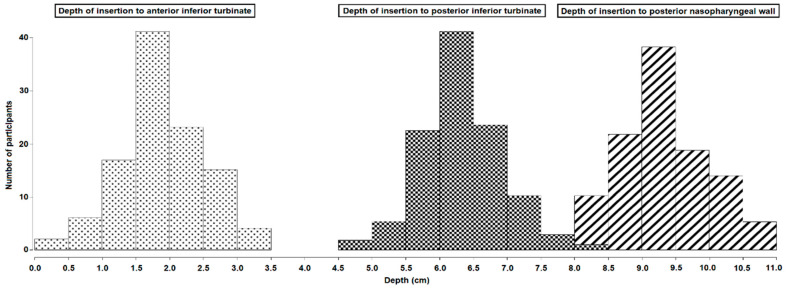
Figure 2.
Depth of insertion to nasal landmarks. Bar charts with the insertion depth to the anterior inferior turbinate (dotted print), posterior inferior turbinate (squared print), and posterior nasopharyngeal wall (striped print). Insertion depth in cm is shown along the x-axis and number of participants along the y-axis.

