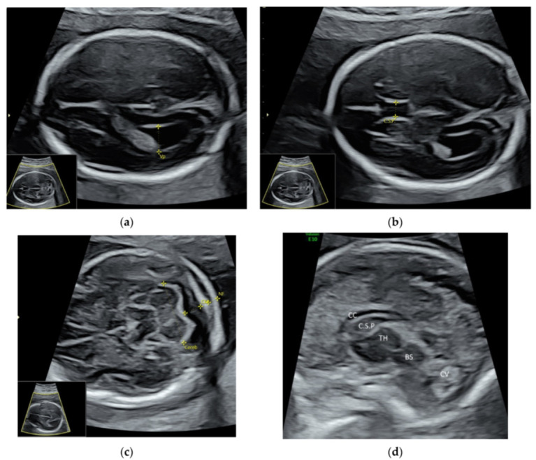
Figure 2.

High-resolution ultrasonography of the fetal brain at 20 weeks’ gestation: transverse views showing (a) posterior horn of the lateral ventricle (Vp), (b) cavum septi pellucidi (C.S.P.), (c) cerebellum (Cereb), Cisterna magna (CM), nuchal fold (NF), and sagittal view showing (d) corpus callosum (CC), thalamus (TH), brain stem (BS), and cerebellar vermis (CV).
