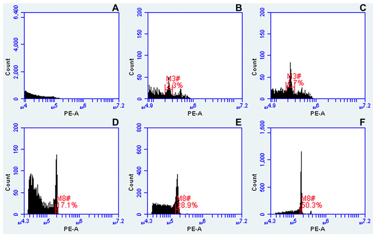
Figure 3.
Comparison of three different standard buffers for nuclei isolation from dried leaves of tetraploid Urochloa accessions, and their effect on histogram quality. (A) Galbraith’s buffer; (B) Otto’s buffer; (C) Otto’s buffer supplemented with 15 mM β-mercaptoethanol and 1% PVP-40 (Modified Otto, see Table 2); (D) Partec buffer; (E) Partec buffer supplemented with β-mercaptoethanol; (F) Partec buffer supplemented with 15 mM β-mercaptoethanol and 1% PVP-40 (Modified Partec, see Table 2). Regions of identification (red marks) were placed across the peaks to export fluorescence values representing peak positions and CVs.

