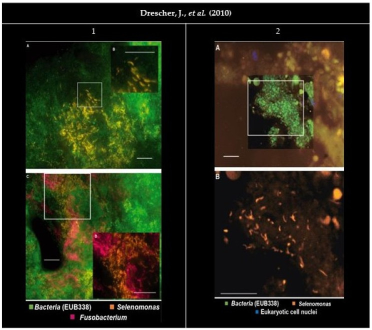
Figure 2.
Panel of the gathered images. (1A,C) Organisms stained by the probe SELE appear as densely packed groups in the cervical portion of the subgingival biofilm. (1B) Higher magnification shows a crescent-shaped morphology. (1C,D) EUB338FITC (Fluorescein isothiocyanate) (green), SELECy3 (Cyanine 3) (bright orange) and FUSOCy5 (Cyanine 5) (magenta). (1D) Higher magnification, with EUB338FITC filter removed for better interpretation. Bars indicate 10 µm. (2) FISH (Fluorescence in situ hybridization) performed on a gingival biopsy. (2A) EUB338FITC (green), SELECy3 (orange), and eukaryotic cell nuclei stained with DAPI (4′, 6-diamino-2-phenylindole) (blue). (2B) Higher magnification took with the Cy3 filter set only. Bars indicate 10 µm. All images were reprinted and adapted with the publisher’s permission.

