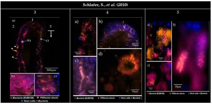
Figure 3.
Panel of the gathered images. (3) Subgingival biofilm visualized by FISH (Fluorescence in situ hybridization). EUB338Cy5 (Cyanine 5) (magenta), FIALCy3 (Cyanine 3) (bright orange), and DAPI (4′, 6-diamino-2-phenylindole) staining (blue). DAPI stains both host cell nuclei and bacteria. (3a1) The carrier tip. (3a2) The carrier side facing the tooth. (3a3) The carrier side facing the pocket epithelium. (3a1,2) Little or no presence of Filifactor alocis. (3a3) Presence of a bright orange signal, indicating a vindicated presence of F. alocis (arrow). The image show artifacts caused by the folding of the embedded carriers (arrowheads). (3b,c) Higher magnification. (3b) Rare colonization of F. alocis amongst the bacteria. (3c) F. alocis in densely packed groups among the organisms on the carrier side facing the soft tissues and host cell nuclei (blue). (4) Establishment of Filifactor alocis in a subgingival biofilm. (4a) Overlay of FIALCy3, EUB338Cy5, and DAPI filter sets. In some parts of the biofilm F. alocis rods can reach a considerable length. (4b,c) Overlay of FIALCy3 and DAPI filter sets. (4b) Radial orientation of F. alocis towards the exterior of a mushroom-like protuberance. (4c) Test-tube-brush formations of F. alocis around signal-free channels. (4d) Overlay of FIALCy3 and EUB338Cy5 filter sets. F. alocis and fusiform bacteria form concentrical structures. (5) Establishment of Filifactor alocis in periodontal tissue. (5a) F. alocis forming tree-like structures among coccoid and fusiform bacteria and autofluorescent erythrocytes. (5b) F. alocis forming palisades with fusiform bacteria around large rod-shaped eubacterial organisms. (5c) F. alocis being part of concentrical bacterial aggregations such as those found in (4d). All images were reprinted and adapted with the publisher’s permission.

