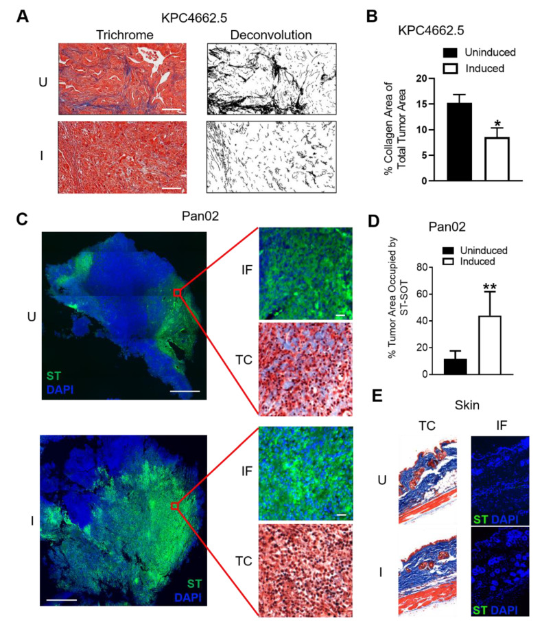
Figure 3.
Induction of ST-SOT leads to intratumoral CN depletion and enhanced ST diffusion in vivo. (A) C57BL/6 mice bearing orthotopic KPC4662.5 tumors (>150 mm3) were administered 5 × 10⁶ colony-forming units (CFU) of ST-SOT by intravenous (i.v.) injection. Forty-eight hours later, groups were intraperitoneally (i.p.) administered PBS (uninduced) or 250 mg L-arabinose (induced) and then euthanized 48 h post-induction. Tumor sections were then subjected to Masson’s trichrome staining (representative images shown, scale bar = 50 µm). Deconvolution of collagen (black) was performed using ImageJ and percent collagen area present within total tumor section (B) was determined. * p < 0.05, t-test. (C) C57BL/6 mice bearing subcutaneous Pan02 tumors (>150 mm3) were i.v. administered 5 × 10⁶ cfu ST-SOT and euthanized 48 h after i.p. injection of PBS (uninduced, U) or L-arabinose (induced, I). Serial tumor sections were subjected to immunofluorescence (IF) staining to detect ST-SOT (ST), as well as trichrome (TC) staining (TC). Representative images shown, scale bar = 200 µm (inset scale bar = 30 µm). Percent area of ST occupying total tumor area (approximated by DAPI staining) (D) was determined using Image One software. ** p < 0.01, t-test. (E) IF and TC staining were performed on skin from Pan02-bearing mice receiving uninduced or induced ST-SOT treatment. Representative images shown.

