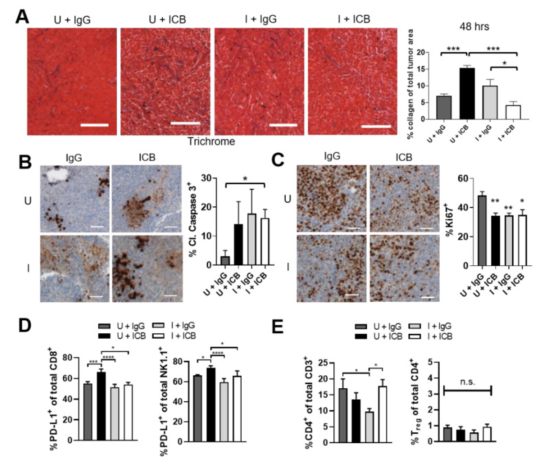
Figure 6.
ST-SOT with immune checkpoint blockade (ICB) treatment augments tumor cell apoptosis, prevents increased expression of PD-L1 associated with ICB, and preserves CD4 populations associated with collagen degradation. C57BL/6 mice bearing s.c. Pan02 tumors (average 100 mm3, n = 3–5) were i.v. administered ST-SOT (5 × 10⁶ CFU) and then i.p. administered 250 mg L-arabinose (induced, I) or PBS (uninduced, U). At induction/PBS, mice were i.p. administered immune checkpoint blockade (ICB) antibodies (anti-PD-1 (200 µg) + anti-CTLA-4 (75 µg)) or control IgG antibody. Forty-eight hours later tumors were excised and processed for sectioning and flow cytometry. (A) Tumors were sectioned, stained using Masson’s trichrome method, and analyzed for collagen density (representative images shown, scale bar = 100 µm). Bar graph represents whole tumor quantification of collagen stain out of total tumor area (n = 3). * p < 0.05, *** p < 0.001, t-test. Sectioned tumors were stained for IHC with anti-cleaved caspase 3 antibody (brown) (B) or anti-Ki67 antibody (brown) (C). Nuclei were counterstained with hematoxylin (blue) (representative images are shown, scale bar = 50 µm). Bar graphs represent whole tumor quantification of positive cells out of total nuclei. * p < 0.05, ** p < 0.01, t-test. (D) Tumors processed for flow cytometry were analyzed for PD-L1-expressing immune subsets and (E) CD4+CD25+FoxP3+ cells (Tregs). * p < 0.05, *** p < 0.001, **** p < 0.0001, t-test.

